 |
Clinical Image
A massive pulmonary embolism with heart strain in urgent care
1 Department of Emergency Medicine, University of Maryland Medical Center, Baltimore, Maryland, USA
Address correspondence to:
Adam C Richardson
DNP, ENP-C, Department of Emergency Medicine, University of Maryland, 110 S Paca Street, 6th Floor, Suite 200, Baltimore, Maryland 21201,
USA
Message to Corresponding Author
Article ID: 101073Z01MB2019
Access full text article on other devices

Access PDF of article on other devices


How to cite this article
Bell ME, Richardson AC. A massive pulmonary embolism with heart strain in urgent care. Int J Case Rep Images 2019;10:101073Z01MB2019.ABSTRACT
No Abstract
Keywords: Computed tomography angiogram, Massive pulmonary embolism, Right heart strain, Saddle pulmonary embolism, Urgent care
Case Report
A 44-year-old male with no significant past medical history self-presented to a community urgent care (UC) complaining of a four day history of shortness of breath, made worse by exertion. Three weeks prior to presentation, the patient underwent arthroscopic surgery on his left knee, and was then sedentary for two weeks following the procedure. He also reported that he travels long distance by car two to three times a week for work, as well as a recent plane trip. His presentation and history made clinicians concerned for pulmonary embolism (PE), so he was transferred to the neighboring emergency department (ED) associated with the UC.
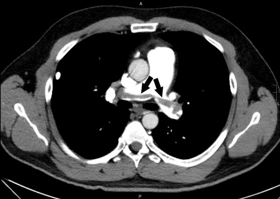
Upon arrival to the ED, his vital signs included blood pressure 116/86 mmHg, pulse 116 bpm, respirations 16, temperature 36.6°C, and oxygen saturation 95% on room air. A D-dimer was obtained and found to be significantly elevated at 9650 ng/mL. A computed tomography angiogram (CTA) of the chest was obtained (Figure 1, Figure 2, Figure 3) which revealed a massive pulmonary thromboembolism with thrombus burden in the bilateral lobar, segmental, and subsegmental branches of all five lobes as well as evidence of right heart strain. A heparin drip was initiated and the patient was admitted to the medical intermediate care service in consultation with the vascular surgery team. A transthoracic echocardiogram revealed a dilated right ventricle with moderately decreased systolic function, septal flattening consistent with right ventricular pressure overload, moderate tricuspid regurgitation, and severe pulmonary hypertension all thought to be secondary to significant clot burden and expected to improve with anticoagulation.


Discussion
A PE is a venous thromboembolism (VTE) in the pulmonary vasculature that occludes blood flow, sometimes leading to sudden death. It is estimated that between 500,000 and 600,000 people in the United States are diagnosed with a PE each year causing 200,000 to 300,000 resultant deaths. Pulmonary embolism is the third leading cause of cardiovascular disease, with an incidence of 1–2 per 1000 individuals [1]. Presenting symptoms of PE most often include tachycardia, tachypnea, dyspnea on exertion, and chest pain. An extensive PE may also have a presentation of syncope, due to low cardiac output or hypotension. The most common risk factors for PE include advanced age (greater than 70 years), prior PEs, malignancy, recent lower limb surgery, chemotherapy, obesity, pregnancy, prolonged immobility, and exogenous estrogen therapy. Computed tomography angiogram is the diagnostic imaging test of choice, but a nuclear medicine ventilation perfusion scan may also be utilized [2]. A saddle PE, now more commonly termed a massive PE is a VTE so large that it straddles the primary pulmonary arterial trunk at the bifurcation which then causes strain on the right side of the heart [3].
Treatment includes immediate anticoagulation, to prevent further incidence. Unfractionated heparin bolus followed by a continuous infusion is the gold standard treatment during hospitalization. Upon discharge from the hospital, oral anticoagulation is typically necessary for approximately 3–6 months while risk factors are addressed and a hypercoagulable workup is completed. In some cases, extended anticoagulation is necessary [2].
Conclusion
This case report highlights the presentation, diagnosis, treatment, and advanced imaging of a man presenting to a UC unaware that he was experiencing a massive PE. A thorough history and physical exam allowed the UC provider to make the important decision to transfer the patient to the ED. Massive PEs are life threatening and cannot miss diagnoses. Pulmonary embolism should be included on the differential diagnosis for any patient presenting with symptoms of chest pain, dyspnea on exertion, tachycardia, and tachypnea, particularly if the patient exhibits any of the aforementioned risk factors.
Laypeople experiencing medical symptoms are expected to essentially self-triage when they decide for themselves whether to present to a primary care office, UC, or ED. While it is widely known that a large portion of the population inappropriately overutilizes the ED for nonemergent medical issues, many people make the opposite mistake and attempt to utilize UC centers with potentially life threatening symptoms because they wish to avoid the lengthy wait times associated with EDs [4]. In this case, UC clinicians quickly identified a patient complaining of shortness of breath as having risk factors for PE and transferred him to the ED where a massive PE with right heart strain was identified by CTA.
REFERENCES
1.
Morrone D, Morrone V. Acute pulmonary embolism: Focus on the clinical picture. Korean Circ J 2018;48(5):365–81. [CrossRef]
[Pubmed]

2.
Lapner ST, Kearon C. Diagnosis and management of pulmonary embolism. BMJ 2013;346:f757. [CrossRef]
[Pubmed]
3.
Alkinj B, Pannu BS, Apala DR, Kotecha A, Kashyap R, Iyer VN. Saddle vs nonsaddle pulmonary embolism: Clinical presentation, hemodynamics, management, and outcomes. Mayo Clin Proc 2017;92(10):1511–8. [CrossRef]
[Pubmed]
4.
May M, Brousseau DC, Nelson DA, et al. Why parents seek care for acute illness in the clinic or the ED: The role of health literacy. Acad Pediatr 2018;18(3):289–96. [CrossRef]
[Pubmed]
SUPPORTING INFORMATION
Author Contributions
Martine E Bell - Conception of the work, Design of the work, Drafting the work, Final approval of the version to be published, Agree to be accountable for all aspects of the work in ensuring that questions related to the accuracy or integrity of any part of the work are appropriately investigated and resolved.
Adam C Richardson - Conception of the work, Design of the work, Drafting the work, Revising the work critically for important intellectual content, Final approval of the version to be published, Agree to be accountable for all aspects of the work in ensuring that questions related to the accuracy or integrity of any part of the work are appropriately investigated and resolved.
Guarantor of SubmissionThe corresponding author is the guarantor of submission.
Source of SupportNone
Consent StatementWritten informed consent was obtained from the patient for publication of this article.
Data AvailabilityAll relevant data are within the paper and its Supporting Information files.
Conflict of InterestAuthors declare no conflict of interest.
Copyright© 2019 Martine E Bell et al. This article is distributed under the terms of Creative Commons Attribution License which permits unrestricted use, distribution and reproduction in any medium provided the original author(s) and original publisher are properly credited. Please see the copyright policy on the journal website for more information.

