 |

|
 |
|
Case Series
| ||||||
| Clinical applications of minimally invasive periodontal plastic surgery | ||||||
| Sangeeta Singh1, Saravanan SP1, Devendra Srivastava1, Raghvendra MH1, A. K. Shreehari1 | ||||||
|
1Department of Dental Surgery, Armed Forces Medical College, Pune.
| ||||||
| ||||||
|
[HTML Abstract]
[PDF Full Text]
[Print This Article]
[Similar article in Pumed] [Similar article in Google Scholar] 
|


| How to cite this article |
| Singh S, Saravanan SP, Srivastava D, Raghvendra MH, Shreehari AK. Clinical applications of minimally invasive periodontal plastic surgery. Int J Case Rep Images 2016;7(12):776–780. |
|
Abstract
|
|
Introduction:
The aim of this paper was to present the excellent results obtained using the principles of minimally invasive surgery in periodontal plastic surgery procedures.
Case Series: This paper will discuss five different procedures: Modified incision design for periodontally accelerated osteogenic orthodontics procedure, papilla reconstruction using the tunnelling approach and connective tissue grafting, pouch and tunnel procedure for adjacent recessions, envelope technique for isolated recession and Liu's Class 1a incision for connective tissue harvesting from the palate. All five techniques resulted in minimal exposure, reduced tissue trauma, minimal postoperative discomfort, better healing and excellent esthetics with perfect tissue color match. Conclusion: The concept of minimally invasive periodontal surgery is especially valid in the field of periodontal plastic surgery resulting in minimal tissue trauma, reduced postoperative discomfort, preservation of the interdental tissues, excellent color match and perfect esthetics. | |
|
Keywords:
Minimally invasive surgery, Periodontally accelerated osteogenic orthodontics reconstruction, Pouch, Tunnel
| |
|
Introduction
| ||||||
|
The concept of minimally invasive surgery has been accepted universally in almost all fields of medicine and has led to the replacement of conventional procedures with minimally invasive ones. The field of periodontology is constantly evolving and reinventing itself at par with the advances in the field of medicine. The introduction of loupes, microscopes and microsurgical instruments has led to the modification of conventional larger access flap designs to the more conservative minimal access approach. This paper will discuss five surgical procedures where the conventional flap designs were modified to allow minimal access for surgery. | ||||||
|
Case Series
| ||||||
|
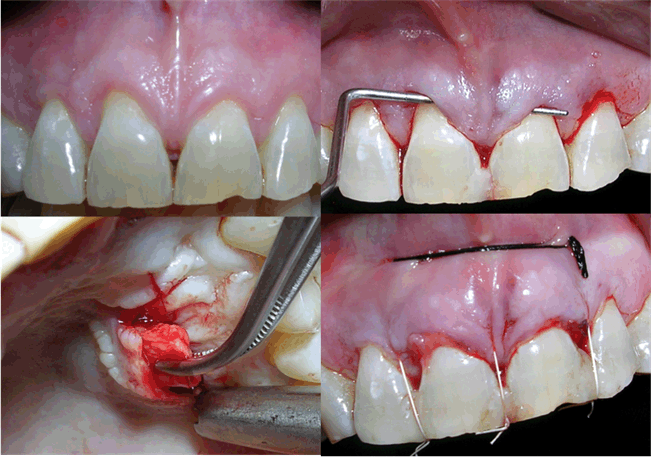
Clinical Scenario Case 2: This patient was very conscious of the dark spaces in her front teeth and came to us seeking correction of the same. She also complained of food lodgement and halitosis. Case 3: This patient was diagnosed with Millers Class II marginal tissue recession in 33, 34. This was associated with sensitivity. Case 4: This young patient was diagnosed with Millers Class III marginal tissue recession. Tension test was positive in 31. He was apprehensive that he will eventually lose the tooth. Clinical management Case 2: Papilla reconstruction using the tunneling approach and connective tissue grafting Case 3: Pouch and tunnel procedure for adjacent recessions Case 4: Envelope technique for isolated recession and Lui's class 1a incision for connective tissue harvesting from the palate Follow-Up | ||||||
| ||||||
| ||||||
| ||||||
| ||||||
| ||||||
|
| ||||||
| ||||||
|
Discussion
| ||||||
|
Mortality and morbidity are unavoidable events of a therapeutic process and are accepted as a part of any therapy. With advances in the field of medicine it has become evident that less invasive methods of interventional treatment in some areas produce far fewer complications with a reduced risk of death and morbidity [6]. Preliminary data from case cohorts and from many studies reveal minimally invasive approach in periodontal surgery has a high potential to seal the healing wound from the contaminated oral environment by achieving and maintaining primary closure. Soft tissues are mostly preserved and minimal gingival recession is observed, an important feature to meet the demands of the patient and the clinician in the aesthetic zone [7]. Modified incision design for periodontally accelerated osteogenic orthodontics procedure Papilla reconstruction using the tunneling approach and connective tissue grafting Pouch and tunnel procedure for adjacent recessions Envelope technique for isolated recession Lui's Class 1a incision for connective tissue harvesting from the palate The modifications in instruments sizes leading to innovations in designs and reduced size has also explored the feasibility of the minimal invasive approach. Research has shown that immune response is better after minimally invasive procedures, leading to better results. | ||||||
|
Conclusion
| ||||||
|
Minimally invasive surgical techniques are now widely accepted and will continue to evolve to further minimize the trauma during surgery. This concept goes a long way in overall patient satisfaction and positive outcomes of any procedure adapted to utilize this technique. | ||||||
|
References
| ||||||
| ||||||


|
[HTML Abstract]
[PDF Full Text]
|
|
Author Contributions:
Sangeeta Singh – Substantial contributions to conception and design, Acquisition of data, Analysis and interpretation of data, Drafting the article, Revising it critically for important intellectual content, Final approval of the version to be published Saravanan SP – Analysis and interpretation of data, Revising it critically for important intellectual content, Final approval of the version to be published Devendra Srivastava – Analysis and interpretation of data, Revising it critically for important intellectual content, Final approval of the version to be published Raghvendra MH – Analysis and interpretation of data, Revising it critically for important intellectual content, Final approval of the version to be published A. K. Shreehari – Analysis and interpretation of data, Revising it critically for important intellectual content, Final approval of the version to be published |
|
Guarantor of submission
The corresponding author is the guarantor of submission. |
|
Source of support
None |
|
Conflict of interest
Authors declare no conflict of interest. |
|
Copyright
© 2016 Sangeeta Singh et al. This article is distributed under the terms of Creative Commons Attribution License which permits unrestricted use, distribution and reproduction in any medium provided the original author(s) and original publisher are properly credited. Please see the copyright policy on the journal website for more information. |
|
|