  |

|
 |
|
Case Report
| ||||||
| An atypical presentation of Lemierre's syndrome with eye and facial pain: A case report with Streptococcus intermedius | ||||||
| Jordana Cheta | ||||||
|
Critical Care and Nephrology Fellow, Eastern Virginia Medical School, 855 W. Brambleton Avenue, Norfolk, VA 23510 United States.
| ||||||
| ||||||
|
[HTML Abstract]
[PDF Full Text]
[Print This Article]
[Similar article in Pumed] [Similar article in Google Scholar] 
|


| How to cite this article |
| Cheta J. An atypical presentation of Lemierre's syndrome with eye and facial pain: A case report with Streptococcus intermedius. Int J Case Rep Images 2016;7(12):848–852. |
|
Abstract
|
|
Introduction:
Lemierre's syndrome is rare but morbid complication that follows an acute oropharyngeal infection. Seeding of an oropharyngeal infection into the lateral pharyngeal space can result in thrombosis of the internal jugular vein with subsequent septic metastasis. The most common presenting symptoms are fever, pharyngitis, odynophagia, oropharyngeal swelling and neck pain. The most common etiologic agent is Fusobacterium necrophorum. We are reporting a rare case in which the causative agent was Streptococcus intermedius and the presenting symptoms were atypical. To the best of our knowledge this is the 4th case of Streptococcus intermedius, resulting from a gingival procedure, with a normal oropharyngeal exam at time of presentation.
Case Report: We describe a 78-year-old Caucasian female presented with eye and facial pain, which resulted to be Lemierre's syndrome. Blood cultures grew Streptococcus intermedius. Patient's hospital course was complicated by parapharyngeal and masticator abscesses. Prompt treatment was initiated and the patient made a full recovery. Conclusion: In the presented case, we had a patient in whom the presenting symptom was mistaken for facial cellulitis resulting in a delay of treatment and progression of symptoms. The presentation was not the typical symptoms of Lemierre's syndrome, such as neck pain, pharyngitis, odynophagia, and fever. Likewise, the blood culture did not grow the usual causative bacteria. Due to the implementation of early treatment, which results in improved outcomes and decreased rates of complications, dependent upon early recognition of the disease, we are reporting this previously undescribed manifestation in hopes to increase awareness across multiple specialties of medicine. | |
|
Keywords:
Atypical Lemierre's presentation, Facial swelling, Streptococcus intermedius infection, Thrombophlebitis of internal jugular vein
| |
|
Introduction
|
|
Lemierre's syndrome, also known as postanginal sepsis, is a rare but potentially life-threatening complication of an acute oropharyngeal infection that results in a septic thrombophlebitis of the internal jugular vein, which can subsequently result in a metastatic infection [1]. In the pre-antibiotic era, Lemierre's syndrome was common and had a mortality rate of 90% [1] . In the recent years the widespread use of antibiotics in acute oropharyngeal infections has resulted in a rapid decline in the prevalence and mortality of Lemierre's syndrome. Lemierre's syndrome is most commonly caused by a gram-negative anaerobic bacillus, Fusobacterium necrophorum (81.7% of cases) [2]. Other organisms that can also result in Lemierre's syndrome are Peptostreptococcus, Group B and C Streptococcus, Staphylococcus, Enterococcus species, and Proteus [2]. Most of these organisms are natural colonizers of the oropharynx. However, when the mucosal barrier is compromised with an acute infection, these agents can enter the blood stream and cause complications such as septic emboli. Baig et al. has described a pattern which exists between the three stages of infection and clinical manifestations in Lemierre's syndrome [1]. In Stage 1, there is an acute oropharyngeal infection, which can present with fever and sore throat. During this stage, the most common presenting symptom (52.2%) is a tender and swollen neck [1]. In Stage 2, the infection invades the pharyngeal space. The onset between Stage 1 and Stage 2 is less than one week [2]. The lateral pharyngeal space is divided by the styloid process into an anterior compartment comprising the sternocleidomastoid muscle, and the posterior neurovascular compartment. Neck pain results from inflammation of the sternocleidomastoid muscle in the anterior compartment. The posterior compartment consists of the carotid artery, internal jugular vein, vagus nerve, lymph nodes, cranial nerves X-XII, and the cervical sympathetic trunk. The clinical findings of invasion of this compartment result from compromise of these structures. During stage 3 septic metastasis to the lungs occur via hematogenous spread [1]. |
|
Case Report
|
|
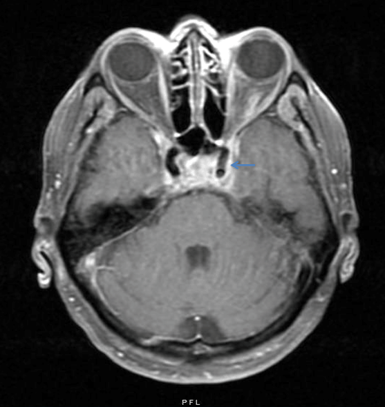
A 78-year-old Caucasian female presented to the emergency department for worsening left sided facial discomfort and new left eye pain. She previously presented to the emergency department three weeks prior with left sided facial discomfort and was diagnosed with facial cellulitis. At that time, she was started on clindamycin, due to an unknown allergy to penicillin, and discharged home. At her second presentation she denied any fever, chills, weight changes, vision loss, neck pain, sore throat, dysphagia, chest pain, palpitations, shortness of breath, nausea, vomiting, and genitourinary symptoms. A computed tomography (CT) scan of the orbits showed significant bilateral internal jugular vein thrombosis with left superior ophthalmic vein involvement. She was started on intravenous clindamycin and piperacillin and tazobactam in the emergency department. Past medical history of the patient was significant for a left lower molar extraction four days prior to the onset of symptoms, as well as hypertension, type II diabetes mellitus and recurrent urinary tract infections. Vital signsof the patient were stable and her physical examination was positive for mild left eye proptosis and chemosis. Extraocular muscles were intact. Swelling and tenderness were present over the left lateral side of the face. Oropharynx examination revealed a well healing extracted left lower molar socket with no erythema, swelling, or exudates. There was no evidence of otitis media, gingivitis, or dental caries. Her neck was non-tender to palpation with no restriction of movement. Cardiopulmonary examination was unremarkable. Laboratory data revealed a white blood cell count of 26.9x10³/mm3 (bands 5%). Liver enzymes were elevated (aspartate aminotransferase 57, alanine aminotransferase 70, and alkaline phosphatase 125). A complete metabolic panel (electrolytes, blood urea nitrogen, creatinine), coagulation profile (prothrombin time, partial thromboplastin time and internationalized normalized ratio), and lactic acid were all within normal range. Electrocardiogram showed sinus rhythm. Blood cultures (4 out of 4 bottles) were positive for Streptococcus intermedius within 48 hours and the organism was found to be susceptible to ampicillin, penicillin, vancomycin and levofloxacin with intermediate sensitivity to clindamycin. Chest radiograph was unremarkable. A computed tomography (CT) scan of the chest with contrast revealed a 7 mm left upper lobe nodule. A CT scan of the neck with contrast upon arrival demonstrated a non-occlusive thrombus in the high cervical segment of the right internal jugular vein, and a left cavernous sinus and ophthalmic vein occlusion. There was no extension into the sigmoid or transverse sinus. (Figure 1). A panorex scan revealed no periodontal disease or frank tooth abscesses. Transthoracic and transesophageal echocardiograms were negative for vegetation. Based on blood culture sensitivities antibiotics were changed to intravenous ceftriaxone and oral metronidazole on day-2. Unfractionated heparin was started due to extensive clot burden. Although the patient was clinically improving with a down trending white blood cell count, on day-7, she developed diplopia and a right lateral rectus nerve palsy. A repeat magnetic resonance imaging (MRI) scan of the head and magnetic resonance venography (MRV) with and without contrast revealed abscesses of the left parapharyngeal, mandibular, and masticator spaces with bilateral partial thrombosis in the cavernous sinus (dorsally on the left (Figure 2) and medially on the right (Figure 3)) with progression of a partial thrombosis in the right internal jugular vein (Figure 4). On day-9 the patient was taken to the operating room by otolaryngology and OMFS for an incision and drainage of the left parapharyngeal and masticator abscesses. On day-12 the patient reported feeling better than she had since admission, was tolerating a diet, and was discharged with home health services on a course of ceftriaxone for four weeks and rivaroxaban for three months. |
|
|
|
|
|
|
|
|
|
Discussion
|
|
Lemierre's syndrome is a rare disease with a high morbidity but low mortality if diagnosed early [3]. It occurs in immunocompetent patients and Fusobacterium necrophorum is the most common causative agent and fever, odynophagia, and pharyngeal inflammation is the most common presentation [3]. To diagnose Lemierre's syndrome imaging and laboratory studies are ordered contingent upon the patient's history, signs, and symptoms. When a patient presents with typical symptoms of pharyngitis, fever, odynophagia, and neck pain the physician is aware to screen for Lemierre's. Laboratory investigations will reveal signs of a bacterial infection with leukocytosis and positive blood cultures. The internal jugular vein thrombosis can be investigated with sonography, CT, or MRI scan-the latter two being more sensitive modalities for identifying thrombosis in intrathoracic and retrosternal veins. Chest X-rays and CT scans of the chest can identify pleural effusions, nodules, cavitation's, infiltrates, and abscesses. Escalona et al. reported a case of a 36-year-old male with Streptococcus intermedius presented with fever and mandibular swelling on examination [3]. Chemlal et al. reported a case of a 21-year-old male with Streptococcus intermedius presented with fever, pharyngitis, and chest pain secondary to pulmonary abscesses [4]. Gupta et al. reported a case of Streptococcus intermedius in which a middle-aged female initially presented with neck pain and persistent headaches (history of dental scraping two weeks prior) but had no fever and a benign oropharyngeal examination. She was discharged with muscle relaxers only to return to the emergency room with worsening of symptoms [2]. All these case reports highlight the importance that a high clinical suspicion and awareness is needed for the diagnosis of Lemierre's syndrome as the presentations are not always typical and can be missed in the early phases of infection. Therapy should be initiated upon suspicion with antibiotics [5]. Routine use of anticoagulation is controversial due to the lack of evidence in literature, but should strongly be considered if there is a clot burden, propagation of clot involving the cavernous sinus, or septic emboli [5]. If there is a lack of improvement with antibiotic therapy, it may be beneficial to drain any abscesses and/or perform ligation of the internal jugular vein in cases where the antibiotic may not be able to penetrate [6] [7]. |
|
Conclusion
|
|
It is important to have Lemierre's syndrome on the differential diagnosis when patients present with facial swelling or eye pain because the manner in which this disease presents itself is not always apparent. As a result, the diagnosis of Lemierre's syndrome can be missed during the early stages of infection. More importantly, a presentation of eye and facial pain should not exclude this diagnosis. Lemierre's syndrome should be considered when patients have a history of dental procedures or odontogenic or oropharyngeal infections and present with worsening symptoms of facial pain. It should be kept in mind that all oral flora could be potential causative agents in this disease. This may prevent a delay in diagnosis, improve outcomes, and prevent severe complications. |
|
References
|
|


|
[HTML Abstract]
[PDF Full Text]
|
|
Author Contributions
Jordana Cheta – Substantial contributions to conception and design, Acquisition of data, Analysis and interpretation of data, Drafting the article, Revising it critically for important intellectual content, Final approval of the version to be published |
|
Guarantor of submission
The corresponding author is the guarantor of submission. |
|
Source of support
None |
|
Conflict of interest
Authors declare no conflict of interest. |
|
Copyright
© 2016 Jordana Cheta. This article is distributed under the terms of Creative Commons Attribution License which permits unrestricted use, distribution and reproduction in any medium provided the original author(s) and original publisher are properly credited. Please see the copyright policy on the journal website for more information. |
|
|