| |
|
|
|
Case Report
| ||||||
| Nigrospora sphaerica causing corneal ulcer in an immunocompetent woman: A case report | ||||||
| Ananya TS1, Anupma Jyoti Kindo2, Anandhalakshmi Subramanian3, Kalpana Suresh4 | ||||||
|
1MBBS, MD Postgraduate, Department of Microbiology, Sri Ramachandra Medical College and Research Institute, Sri Ramachandra University, Chennai, Tamil Nadu, India.
2MD, Professor, Department of Microbiology, Sri Ramachandra Medical College and Research Institute, Sri Ramachandra University, Chennai, Tamil Nadu, India. 3MBBS, MS Postgraduate, Department of Ophthalmology, Sri Ramachandra Medical College and Research Institute, Sri Ramachandra University, Chennai, Tamil Nadu, India. 4MS, Professor, Department of Ophthalmology, Sri Ramachandra Medical College and Research Institute, Sri Ramachandra University, Chennai, Tamil Nadu, India. | ||||||
| ||||||
|
[HTML Abstract]
[PDF Full Text]
[Print This Article]
[Similar article in Pumed] [Similar article in Google Scholar] 
|
| How to cite this article |
| Ananya TS, Kindo AJ, Subramanian A, Suresh K. Nigrospora sphaerica causing corneal ulcer in an immunocompetent woman: A case report. Int J Case Rep Images 2014;5(10):675–679. |
|
Abstract
|
|
Introduction:
Corneal ulcers secondary to trauma can often pose a clinical challenge. The causative pathogen is at times an unusual and resistant microorganism which may not be identified by conventional laboratory techniques.
Case Report: A 45-year-old immunocompetent woman with a history of injury of right eye with a cow's tail was diagnosed to have fungal corneal ulcer. Sporulation of the isolate occurred after prolonged incubation and the pathogen was found to be Nigrospora sphaerica. The number of cases of true infection caused by this fungus amount to only five in available literature. The patient did not improve with medical management using natamycin and ketoconazole and underwent voriconazole therapy. Conclusion: Delayed sporulation of pathogenic fungal isolates may necessitate prolonged incubation and use of multiple sporulation techniques for the purpose of speciation. Uncommon fungi also need to be kept in mind when dealing with an unresponsive/worsening clinical situation. | |
|
Keywords:
Fungal keratitis, Nigrospora sphaerica, Black fungi, Voriconazole
| |
|
Introduction
| ||||||
|
Nigrospora species are a filamentous melanized (dematiaceous) group of fungi that belong to the phylum Ascomycota [1]. Widespread in the environment and every so often found in the lab as a contaminant, it is only rarely encountered as a true pathogen. Thus far, literature includes only five cases of human infections caused by this violently spore discharging fungus [2], which includes infections of the skin, nail and eye in both immunocompetent and immunocompromised patients [3] [4] [5] [6] [7]. Herein, we report another incidence of this fungus exhibiting its infrequent pathogenic role. | ||||||
|
Case Report
| ||||||
|
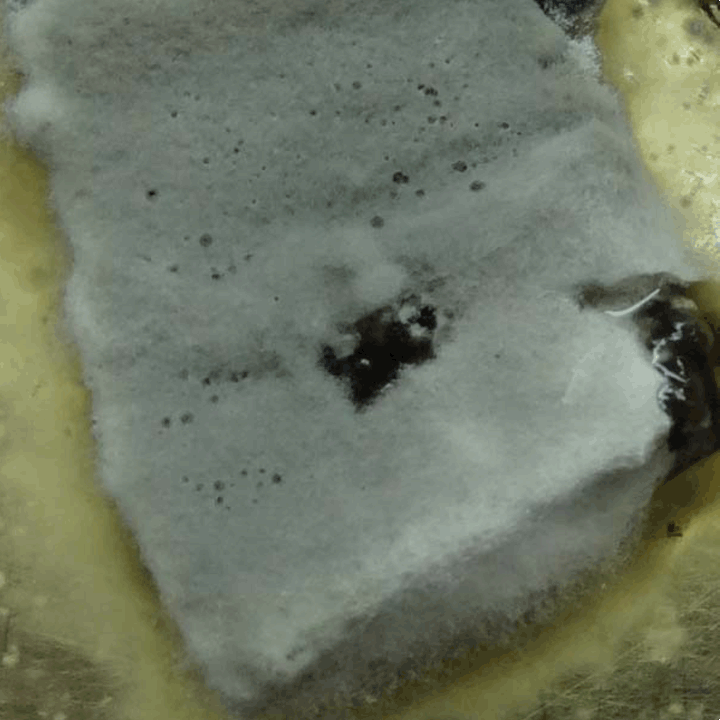
A 45-year-old previously healthy immunocompetent woman hailing from a rural area in south India presented to the ophthalmology outpatient department of our institute on September 2013 with a history of injury of the right eye following a hit from a cow's tail one month before. She had complaints of redness, pain and watering of the right eye since last one month, associated with decrease in vision which was gradual in onset, beginning after two days of the injury and progressive in nature. She was not a diabetic and was not on any medication. Examination of the affected eye revealed the visual acuity to be limited to counting fingers at one meter, with a congested anterior chamber and corneal infiltration with extensive surrounding stromal edema in the left lower quadrant (Figure 1). Gram stain and 10% KOH mount of the corneal scrapings were sent to the microbiology lab along with the inoculated culture plates of Sheep Blood Agar, Chocolate Agar and Sabouraud's Dextrose Agar without cycloheximide (HiMedia Laboratories, Mumbai, India). The Gram stain showed the presence of occasional inflammatory cells but no microorganisms while the 10% KOH mount revealed the presence of septate hyaline hyphal elements. The culture plates were incubated at 25°C. In seven days, the plates showed moldy growth along the sickle shaped stab lines of inoculation. The colonies were woolly in texture, with the color turning from white to grey to black (Figure 2), producing a reverse pigmentation which darkened from pale to dark brown and finally to black (Figure 3). A slide culture using Oatmeal Agar (HiMedia Laboratories, Mumbai, India) was put up for the purpose of speciation and incubated at room temperature. An LPCB (Lactophenol Cotton Blue) mount of the slide culture ten days later demonstrated only sterile hyphae and no spores. In an attempt to induce sporulation, a banana peel culture was put up, which has been indigenously developed in our lab (Figure 4). Banana peel was cut up into smaller pieces of one square inch and autoclaved in a glass Petri dish. No agar needs to be added since the banana peel itself acts as the source of nutrition. The fungal isolate was inoculated onto the sterile pieces, moistened with a few drops of sterile distilled water and incubated at room temperature. The banana peel simulates the saprophytic environment. Examination with an LPCB mount three weeks later revealed branching septate hyphae, with the conidiophores exhibiting a single conidium at their inflated apex (Figure 5). The conidia were unicellular, very black, ovoid in shape, with the older spores showing a horizontal flattening (Figure 6). The description was found to fit the characteristic "black spores" of Nigrospora sphaerica. The patient was initially managed medically with oral ketoconazole and natamycin eye drops after the diagnosis of fungal keratitis was made but showed no improvement. Further to this, bandage contact lens was used for the descemetocele. A month after presentation to the outpatient department, she was started on oral voriconazole 400 mg per day for 10 days and 1% voriconazole eye drops two hourly (eight times a day) for 28 days. The patient showed full recovery on subsequent follow-up visits with reduction in stromal oedema, redness and clearing of corneal infiltration leading to full recovery of vision (6/9 with refraction) (Figure 1). | ||||||
|
| ||||||
| ||||||
| ||||||
| ||||||
| ||||||
| ||||||
|
Discussion
| ||||||
|
Spores of Nigrospora species are more abundant in regions with a tropical or subtropical climate, more commonly occurring outdoors than indoors and readily isolated from soil and decaying plants [8]. It is a frequent plant pathogen affecting grain and fruit [7, 8]. Our patient suffered a traumatic inoculation of the spores on being hit with a cow's tail most likely contaminated with soil and vegetative matter which are constant habitats of this widely distributed fungus. Of the seven species of Nigrospora that are known, N. oryzae and N. sphaerica are the more frequently encountered, with the former being a sizeable agricultural problem. The usual human response to this fungus is hay fever or asthma [8] but singular cases of infections have been reported, with N. sphaerica being the pathogen and trauma being associated with the infections in the immunocompetent [3] [4] [5] [6] [7] . Nigrospora has also been isolated as part of the conjunctival flora in a healthy conjunctival sac [9]. A study of the frequency of occurrence of the spores of Nigrospora species at every month of the year in the United States [8], revealed that Nigrospora spores are typically present outdoors throughout the whole year at a consistent but low density but tend to be higher from August to October. Although there is no data available on the spore density of Nigrospora in India, it is of interest to note that our patient suffered her injury during the month of August. In culture, the color of the colony darkens in proportion with the increasing amount of sporulation during incubation [10]. A full grown culture was black on both the obverse and reverse sides. The asexual spores (conidia) were found to be typical of the textbook description with conidia ranging from young ovoid to older flattened [8]. The teleomorphic stage (sexual phase) of Nigrospora are included in Khuskia, another genus of the phylum Ascomycota. The fungal isolate has been deposited at The Centraalbureau voor Schimmelcultures (CBS) Fungal Biodiversity Centre, The Netherlands, and can be accessed with the accession number CBS 137557. | ||||||
|
Conclusion
| ||||||
|
The necessity of considering rarer, more resistant pathogens when managing a case of fungal keratitis is emphasized in this case, since our patient showed clinical improvement only after changeover to voriconazole therapy. Although Nigrospora sphaerica grew very rapidly in culture of the clinical specimen, sporulation took nearly five weeks which would warrant prolonged incubation and usage of multiple techniques like slide culture and banana peel culture for the purpose of accurate speciation of those pathogenic fungi showing no or delayed sporulation. | ||||||
|
Acknowledgements
| ||||||
|
We acknowledge The Centraalbureau voor Schimmelcultures (CBS) Fungal Biodiversity Centre, The Netherlands, for depositing our clinical isolate in the CBS Collection of Fungi. | ||||||
|
References
| ||||||
| ||||||
|
[HTML Abstract]
[PDF Full Text]
|
|
Author Contributions
Ananya TS – Acquisition of data, Analysis and interpretation of data, Drafting the article, Critical revision of the article, Final approval of the version to be published Anupma Jyoti Kindo – Conception and design, Acquisition of data, Analysis and interpretation of data, Drafting the article, Critical revision of the article, Final approval of the version to be published Anandhalakshmi Subramanian – Acquisition of data, Critical revision of the article, Final approval of the version to be published Kalpana Suresh – Conception and design, Acquisition of data, Analysis and interpretation of data, Critical revision of the article, Final approval of the version to be published |
|
Guarantor of submission
The corresponding author is the guarantor of submission. |
|
Source of support
None |
|
Conflict of interest
Authors declare no conflict of interest. |
|
Copyright
© 2014 Ananya TS et al. This article is distributed under the terms of Creative Commons Attribution License which permits unrestricted use, distribution and reproduction in any medium provided the original author(s) and original publisher are properly credited. Please see the copyright policy on the journal website for more information. |
|
|
|
About The Authors
| |||
| |||
| |||
| |||
| |||