| Table of Contents |  |
|
Case Report
|
| Bilaterally symmetrical involvement in hirayama disease: A rare presentation |
| Anand Pai1, Umadevi V2, Suresh Saravanakumar3 |
|
1MBBS, Resident Doctor, Department of General Medicine, Aarupadai Veedu Medical College and Hospital, Pondicherry, India.
2MD, Professor, Department of General Medicine, Aarupadai Veedu Medical College and Hospital, Pondicherry, India. 3MD, Associate Professor, Department of General Medicine, Aarupadai Veedu Medical College and Hospital, Pondicherry, India. |
|
doi:10.5348/ijcri-2012-08-165-CR-12
|
|
Address correspondence to: Anand Pai Department of General Medicine Aarupadai Veedu Medical College and Hospital Kirumampakkam, Pondicherry- 607402 India Phone: Mob: +919841320712; Ph: 044-28342171 Email: cdrocks_87@yahoo.co.in |
|
[HTML Abstract]
[PDF Full Text]
|
| How to cite this article: |
| Pai A, Umadevi V, Saravanakumar S. Bilaterally symmetrical involvement in hirayama disease: A rare presentation. International Journal of Case Reports and Images 2012;3(8):43–45. |
|
Abstract
|
|
Introduction:
Hirayama disease (HD) or otherwise flexion cervical myelopathy is characterized by progressive muscular weakness and atrophy of the distal upper limbs most frequently seen in young males. Hirayama disease is thought to be secondary to an abnormal anterior displacement of the posterior dura with secondary compression of the lower cervical spinal cord and chronic injury to the anterior grey horn cells of cervical cord leading to localised cord atrophy. HD (brachial monomelic amyotrophy) is a unilateral or grossly asymmetric bilateral disease.
Case Report: A 26-year-old male who presented with progressive weakness and wasting in the distal muscles of both the upper limbs with no sensory involvement and autonomic involvement. Clinical, radiological and electrophysiologic findings in our patient were consistent with the diagnosis of HD. The patient was advised to wear a soft cervical collar. On three month follow up the patient had shown no progression of his disease. Conclusion: There are very sparse reports of bilaterally symmetric involvement and hence we report this case as bilateral symmetric is a severe form of the classic disease which remains undiagnosed due to a common notion that it is a unilateral or grossly asymmetric disease, hence we report this case. | |
|
Key Words:
Hirayama disease, Flexion cervical myelopathy, Bilateral symmetric involvement
| |
|
Introduction
| ||||||
|
Hirayama disease, a rare neurological disease, is characterized by insidious unilateral or bilateral muscular atrophy and weakness of the forearms and hands, without sensory or pyramidal signs. [1] The disease primarily affects men in the second to third decades. The disease progresses initially, but is followed by spontaneous arrest after few years, unlike motor neuron disease which has a steady progression. [2] Hirayama disease is characterized by focal ischemic changes in the anterior horn cells of the lower cervical cord that result in amyotrophy, which is usually unilateral but may also be bilateral. There are very sparse reports hence we present a case with HD and discuss its pathophysiology and imaging characteristics. | ||||||
|
Case Report
| ||||||
|
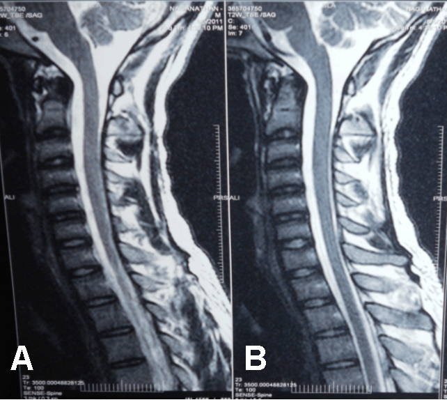
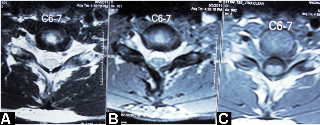
A 26-year-old male presented with progressive weakness and wasting in the distal muscles of both the upper limbs for last seven months. He noticed weakness while lifting dumbells, and later developed difficulty in handling small utensils and writing. He had no sensory symptoms. There was no preceding illness, trauma, exposure to toxins. His past medical history was noncontributory; he denied any allergic disease and none of his family members had similar symptoms. Neurological examination showed significant muscle loss of both upper limbs involving all muscle groups except for the deltoid and brachioradialis muscles. Strength was reduced in the finger extensors, abductor digit mini, flexor pollicis longus, abductor pollicis brevis, pronator teres and ulnar innervated muscles. Strength in lower limbs muscles was normal. Deep tendon reflexes were normal except slight hyporeflexia in triceps, the knee jerks were brisk bilaterally. Plantars were flexors. Mild action tremor was seen in fingers. Higher mental functions, gait, cranial nerves, cerebellar functions, and sensory examination were normal. There was no evidence of polyminimyoclonus or autonomic disturbances. The hematological investigations were normal. Cerebrospinal fluid showed glucose: 52 mg/dL, proteins: 13 mg/dL, and white cells: 4/mm3. Serum IgE was within normal limits. Electrophysiologic studies showed decreased amplitude in ulnar nerves with normal latencies and borderline low velocities; giant motor unit action potentials were seen in the muscles innervated by C7, C8 and T1 nerve roots, with fibrillations and positive waves. There was no evidence of conduction block. Plain cervical spine radiograph showed no abnormality and no misalignment of vertebral bodies. The magnetic resonance imaging (MRI) of the cervical spine in neutral position showed mild atrophy with mild loss of attachment between posterior dural sac and subjacent lamina of cervical cord at C6–C7 (Figure 1). On flexion view MRI cervical spine, there was anterior displacement of the dural sac and spinal cord with widening of posterior epidural space with engorgement of epidural veins (Figure 2). The axial sections showed anteroposterior flattening of the cord with intrinsic signal changes (Figure 3). Clinical, radiological and electrophysiologic findings in our patient were consistent with the diagnosis of bilateral symmetric Hirayama disease. The patient was advised to wear a soft cervical collar. On a three-month follow up the patient had shown no progression of his disease. | ||||||
|
| ||||||
|
| ||||||
| ||||||
|
Discussion
| ||||||
|
This disease was described first by, and named after, Hirayama in 1959 and most cases of this disease have been reported from Japan and India. [3] This non-familial disorder mostly hits young men, progresses slowly and is often self-limited. Researchers argue that wasting and weakness associated with this disease is because of imbalanced growth between the patient's vertebral column and spinal canal contents. [4] The imbalanced growth causes a tight dural sac and displaces posterior dural wall anteriorly when the neck is flexed. Normally, the spinal dura mater is loosely anchored to the vertebral canal by the nerve roots and the periosteum at the foramen magnum and the dorsal surfaces of C2 and C3, and the other at the coccyx. The dura mater is slack enough to adjust to the increased length of cervical spine in flexion. In patients with Hirayama disease, however, the tight dural sac separates the posterior dural sac from its subjacent lamina and on neck flexion, cannot compensate for the increased length of the wall. The posterior dural wall shifts anteriorly, and the cervical spinal cord gets compressed against the posterior margin of adjacent vertebral bodies. This repetitive compression affects the anterior spinal artery and causes microcirculatory disturbances in its territory in the lower cervical cord; [1] the anterior horn cells which are vulnerable to ischemia begin to degenerate, resulting in localized cord atrophy of the lower cervical region which proves to be evident in our case. Hirayama disease is also associated with raised levels of serum IgE and several allergic disorders. Blood often stagnates in the compressed lower cervical cord allowing platelets to aggregate and cause histamine to be released, factors that cause arterial spasm and microcirculatory disturbances. [5] Moreover, the finding that atopic disorders affect young people more than the elderly, men more than women, and Asians more than non-Asians may in part explain the distribution of Hirayama disease. In young males with distal upper extremity weakness, atrophy, preserved reflexes and normal sensory examination, Hirayama disease should be considered. Initial unilateral involvement or bilateral involvement, unilateral or bilateral tremor, absence of cerebellar and sphincter involvement, and EMG findings of anterior horn cell disease further support the diagnosis. Syringomyelia, spinal cord tumors, poliomyelitis, multifocal motor neuropathy, and toxic neuropathies should be excluded. A cervical MRI with neck flexed can pick up dynamic cord compression if a neutral position MRI fails to demonstrate the classical findings which was done in our case.[6] | ||||||
|
Conclusion
| ||||||
|
Bilaterally symmetric Hirayama disease is a severe form of a classic disease which remains undiagnosed due to a common notion that it is a unilateral or grossly asymmetric disease. This description calls for review of the term "brachial monomelic amyotrophy" described to denote this disease. Hirayama's disease has a self-remitting course over a few years and therefore the treatment involves reducing repeated trauma to the cervical cord by use of a cervical collar [6]. Early recognition of this disease is necessary because limitation of neck flexion can prevent further progression of this disease. | ||||||
|
References
| ||||||
| ||||||


|
[HTML Abstract]
[PDF Full Text]
|
|
Author Contributions:
Anand Pai – Conception and design, Acquisition of data, Analysis and interpretation of data, Drafting the article, Critical revision of the article, Final approval of the version to be published Umadevi V – Conception and design, Analysis and interpretation of data, Critical revision of the article; Final approval of the version to be published Suresh Saravankumar – Analysis and interpretation of data, Critical revision of the article, Final approval of the version to be published |
|
Guarantor of submission:
The corresponding author is the guarantor of submission. |
|
Source of support:
None |
|
Conflict of interest:
Authors declare no conflict of interest. |
|
Copyright:
© Anand Pai et al. 2012; This article is distributed the terms of Creative Commons Attribution License which permits unrestricted use, distribution and reproduction in any means provided the original authors and original publisher are properly credited. (Please see Copyright Policy for more information.) |
|
|